• Skip to primary navigation
  • Skip to main content
PATHS-UP

PATHS-UP

Precise Advanced Technologies and Health Systems for Underserved Populations

MENUMENU
  • About
        • About
        • Contact Us
        • Job & Research Board
  • People
        • People
        • PATHS-UP Faculty & Staff
        • Student Leadership Council
        • Paths-Up Leadership Team
        • Advisory Boards
          • Dean’s Council
          • Scientific Advisory Board
          • Workforce Development Advisory Board
  • News
  • Research
        • Research
        • Amplification biochips and implantable bar-code sensors | Thrust 1
          • T1 Publications
        • Mobile computational imaging and spectroscopy systems | Thrust 2
          • T2 Publications
        • Wearable sensing and imaging technologies | Thrust 3
          • T3 Publications
        • Remote bio-behavioral inference and patient response | Thrust 4
          • T4 Publications
        • Milestones
  • Industry
        • Industry
        • Join Us
        • Partners
        • Innovation Seed Fund Program
        • PATHS-UP: SBIR/STTR and ERC Collaboration Opportunity (SECO)
        • Innovation Ecosystem Team
        • 2023 PATHS-UP Digital Health Workshop
          • 2023 Digital Health Workshop Seed Fund Winners
          • 2023 PATHS-UP Digital Health Workshop in Review
        • 2024 PATHS-UP Digital Health Workshop
  • Workforce Development
    • Workforce Development
      • Professional Development Activities
    • K-12
      • Research Experiences for Teachers (RET)
      • Young Scholars Program
    • Undergraduates
      • PATHS-UP Scholar Expectations
      • BS Engineering/Masters of Public Health Program
      • Vertically Integrated Projects (VIP) Program
      • Summer Research Experience for Undergraduates (REU)
    • Graduates
      • Experiential Learning Program for Graduate Students
      • Fellowship Funding Opportunities
  • Member Portal

Thrust 3

You are here: Home / Research / Thrust 3

Wearable Sensing and Imaging Technologies

Thrust Leads

FIU (Thrust Co-Lead)

Jessica Ramella-Roman

FIU (Thrust Co-Lead)
Rice
(Thrust Co-Lead)

Ashok Veeraraghavan

Rice (Thrust Co-Lead)

Goal: Thrust 3 will develop wearable devices for blood pressure, heart rate and heart rate variability monitoring and readers for the implantable bar-code sensors for point-of-care (POC) monitoring of cardiovascular disease and diabetes biomarkers. In addition, T3 is developing realistic computational models in support of all thrusts that account for patient-specific anatomy and physiology including skin tone and elevated Body Mass Index.

Project 3.1: Enabling wearable technologies for individuals experiencing disability within the target PATHS-UP communities

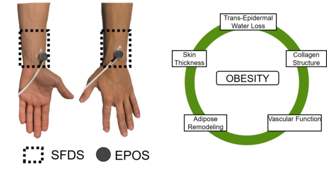
Obesity afflicts a large portion of the PATHS-UP target population. Through computational work, we have demonstrated that the combination of obesity and elevated skin tone has dramatic effects on optical signals such as the ones found in photoplethysmography (PPG). Our goal in this project is to focus on the unique challenges of the obese population within and refine the PATHS-UP wearable technologies through a combination of computational modeling and human studies aimed at characterizing the skin of the obese.
FIU (Thrust Co-Lead)

Jessica Ramella-Roman

FIU (Thrust Co-Lead)
EPOS image

THRUST 3.1 RESEARCH HIGHLIGHTS

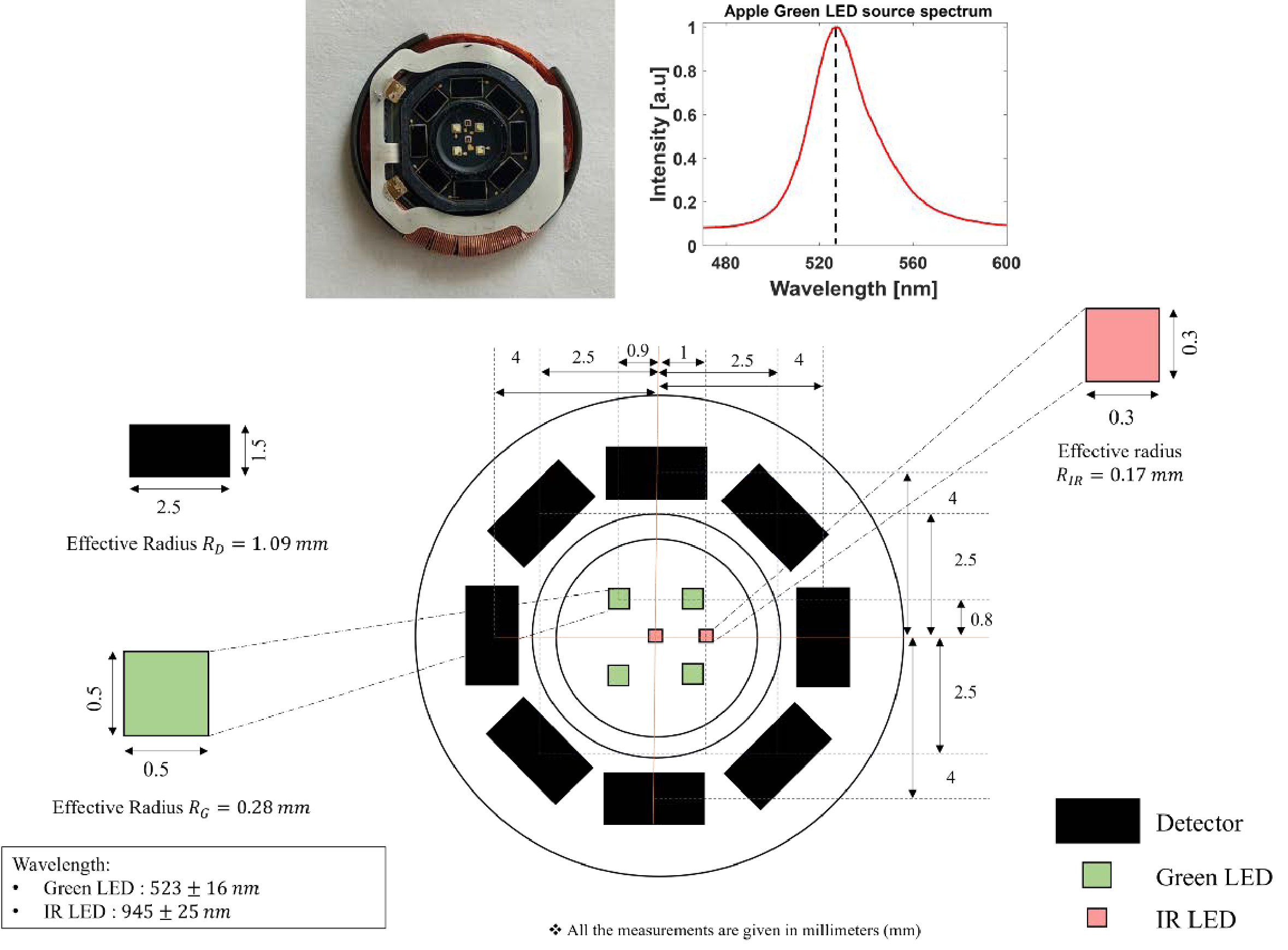
Fig. 1. Schematic of the heart rate sensor specifications in Apple Watch S5.

Biomedical Optics Express (2021)

Monte Carlo analysis of optical heart rate sensors in commercial wearables: the effect of skin tone and obesity on the photoplethysmography (PPG) signal

Ajmal, Tananant Boonya-Ananta, Andres J. Rodriguez, V. N. Du Le, and Jessica C. Ramella-Roman
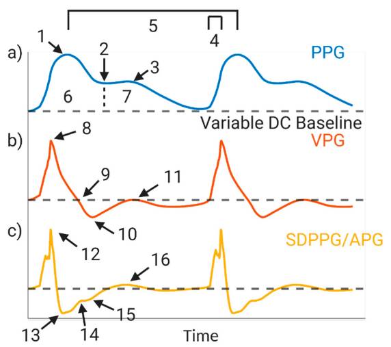
a) Photoplethysmography (PPG) waveform: 1. systolic peak; 2. dicrotic notch; 3. diastolic peak; 4. slope transit time; 5. heart rate; 6. area under systolic waveform; 7. area under diastolic waveform; (b) first derivative, velocity plethysmograph (VPG): 8. max slope in systole; 9. end of systolic peak; 10. Start of dicrotic notch; 11. max slope in diastole; (c) second derivative, second derivative photoplethysmograph or acceleration plethysmograph (SDPPG/APG): 12. a-point; 13. b-point; 14. c-point; 15. d-point; 16. e-point. Created with BioRender.com.

Biosensors (2021)

Sources of Inaccuracy in Photoplethysmography for Continuous Cardiovascular Monitoring

Jesse Fine, Kimberly L. Branan, Andres J. Rodriguez, Tananant Boonya-ananta, Ajmal, Jessica C. Ramella-Roman, Michael J. McShane and Gerard L. Coté

Project 3.2: Multi-modal physiological wearable sensing platform

This project aims to develop a multimodal physiological sensing wearable device that will be placed along the radial artery and will be used to measure heart rate, heart rate variability, and cuffless blood pressure.

TAMU

Peggy Wang

TAMU
TAMU

Gerard Cote

TAMU
Rice
(Thrust Co-Lead)

Ashok Veeraraghavan

Rice (Thrust Co-Lead)
TAMU

Limei Tian

TAMU
FIU

Nezih Pala

FIU
multimodal physiological sensing wearable device that will be placed along the radial artery and will be used to measure heart rate, heart rate variability, and cuffless blood pressure.

THRUST 3.2 RESEARCH HIGHLIGHTS
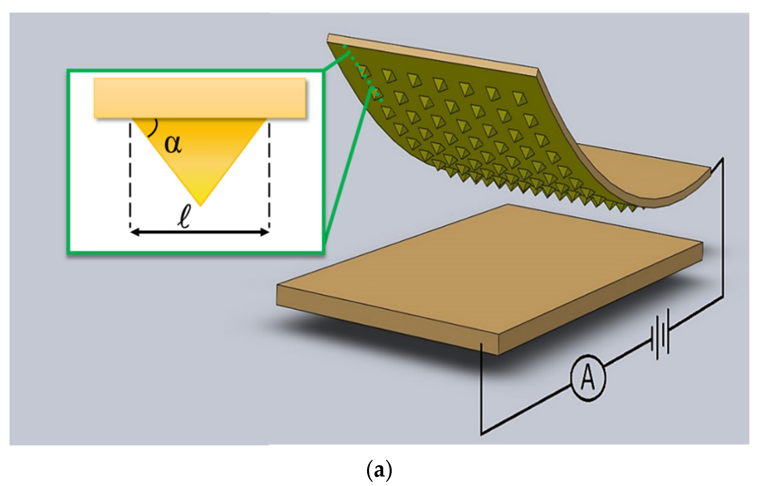
Figure 1. Piezo-resistive sensor. (a) Schematic of a micro-pyramid piezo-resistive sensor showing parameters such as pyramid angle “α” and pyramid base size “ℓ”.

Micromachines (2022)

Design Rules for a Wearable Micro-Fabricated Piezo-Resistive Pressure Sensor

Borzooye Jafarizadeh, Azmal Huda Chowdhury, Iman Khakpour, Nezih Pala and Chunlei Wang
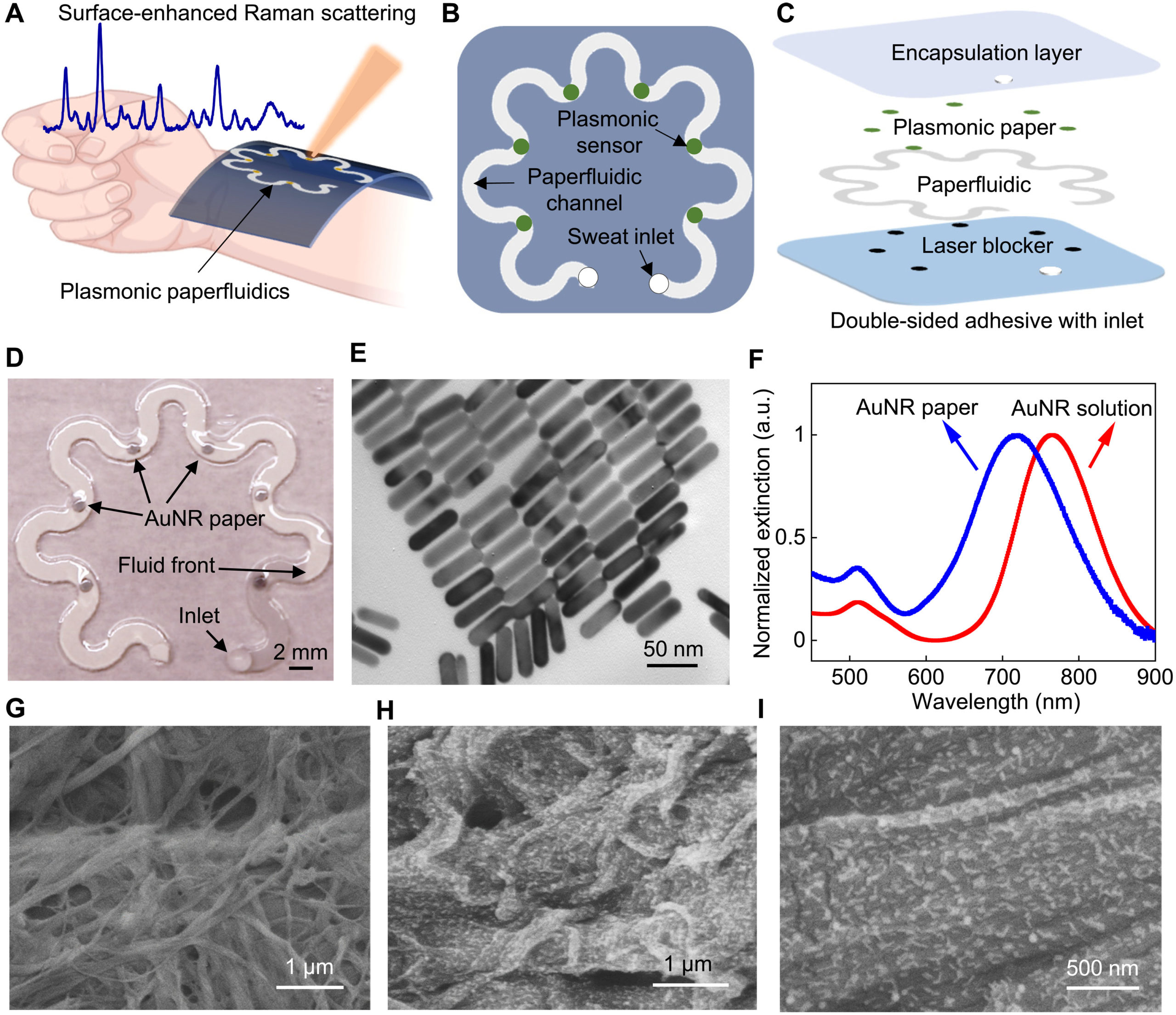
Fig. 1. Wearable plasmonic paper microfluidic device. (A) Conceptual illustration of a wearable plasmonic paperfluidic device for sweat collection, storage, and in situ analysis using SERS. (B) Top view and (C) stacked view schematics of the paperfluidic device highlighting key functional layers. (D) Photograph of an assembled paperfluidic device with six plasmonic sensors. (E) TEM image of gold nanorods (AuNRs) with a uniform size distribution. (F) Extinction spectra of AuNR solution and AuNR paper. a.u., arbitrary units. SEM images of pristine chromatography paper (G) and AuNR paper (H and I).

Science Advances (2022)

Wearable plasmonic paper–based microfluidics for continuous sweat analysis 

Umesha Mogera, Heng Guo, Myeong Namkoong, Md Saifur Rahman, Tan Nguyen, Limei Tian
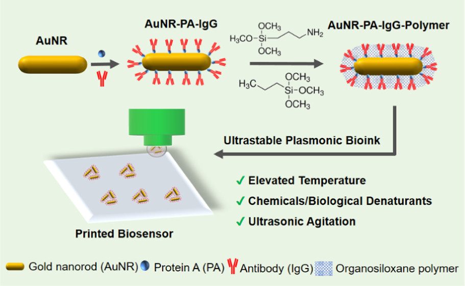
Schematic illustration showing the preparation steps for the printable plasmonic bioink and biochip, including AuNR–PA–IgG bioconjugation, in situ polymerization, and bioink printing. The printed plasmonic biochip is thermally, chemically, and mechanically stable under various harsh conditions.

ACS Applied Materials & Interfaces (2021)

Ultrastable Plasmonic Bioink for Printable Point-Of-Care Biosensors 

Ze Yin, Heng Guo, Yixuan Li, Joshua Chiu, and Limei Tian
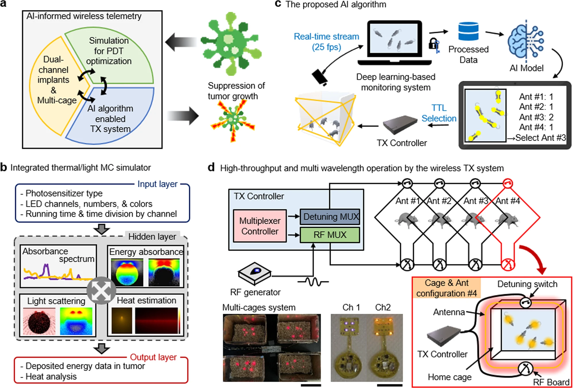
a Overview of the proposed integrated platform. b Workflow for an integrated MC simulator for analysis of heat dissipation and light propagation. c The proposed AI-based algorithm for real-time monitoring of mice. d Schematic illustration of the low-power wireless telemetry system for multi-wavelength operation (top). Two images represent demonstration of multichannel activation using a single power source (bottom, left) and multi-wavelength operation (bottom, right); scale bar 10 cm (left) and 1 cm (right).

Nature Communications (2022

AI-enabled, implantable, multichannel wireless telemetry for photodynamic therapy

Woo Seok Kim, M. Ibrahim Khot, Hyun-Myung Woo, Sungcheol Hong, Dong-Hyun Baek, Thomas Maisey, Brandon Daniels, P. Louise Coletta, Byung-Jun Yoon, David G. Jayne & Sung Il Park 

Project 3.3: Wearable high resolution optical imagers

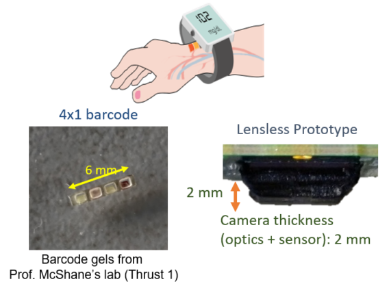
This project focuses on designing and building a wearable imager with integrated illumination for measuring vital information, such as glucose and oxygen content in blood, through the skin tissue. The vital information will be measured via the response of an implanted barcode that is being developed in Thrust 1. The wearable imager is designed to image the barcode through tissue scattering with sufficient spatial resolution and will have controlled illumination to activate and record the phosphorescence lifetime response of the barcode.

FIU (Thrust Co-Lead)

Jessica Ramella-Roman

FIU (Thrust Co-Lead)
Rice
(Thrust Co-Lead)

Ashok Veeraraghavan

Rice (Thrust Co-Lead)
UCLA

Aydogan Ozcan

UCLA
Wearable imager with lensless camera

THRUST 3.3 RESEARCH HIGHLIGHTS

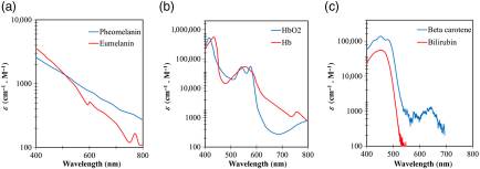
Extinction coefficient spectrum for major absorbers present in human skin: (a) pheomelanin and eumelanin, (b) oxygenated hemoglobin (HbO2) and deoxygenated hemoglobin (Hb), and (c) beta carotene and bilirubin from Ref. 22.

Journal of Biomedical Optics (2022)

Skin optical properties in the obese and their relation to body mass index: a review 

Andres J Rodriguez, Mel Tananant Boonya-Ananta, Mariacarla Gonzalez, Vinh Nguyen Du Le, Jesse Fine, Cristina Palacios, Mike J McShane, Gerard L Coté, Jessica C Ramella-Roman 
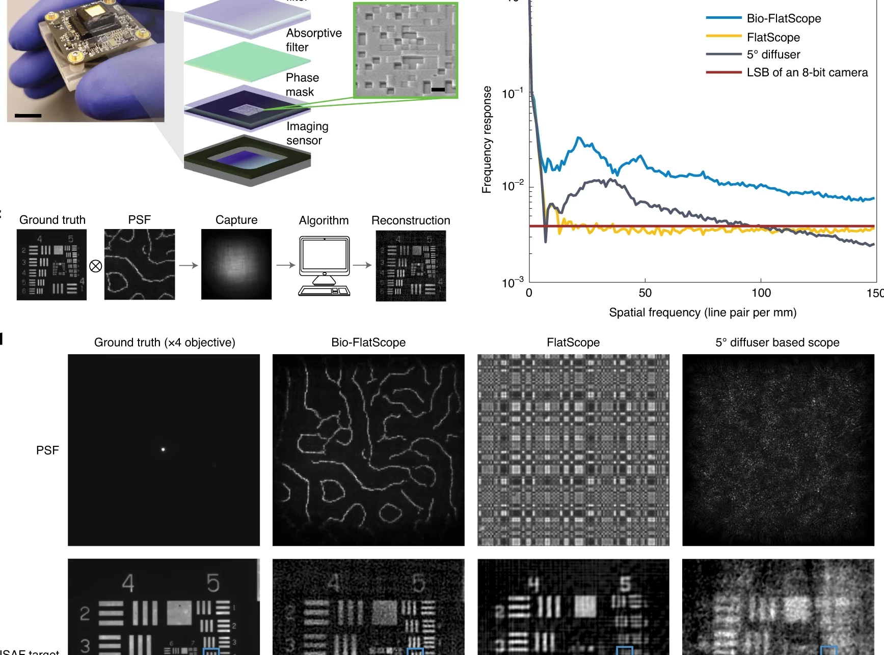
Photo of a Bio-FlatScope prototype including the off-the-shelf board level camera. Scale bar, 10 mm. Zoom-in shows components that filter excitation light and apply the phase mask transfer function to the incident light. Also shown is a scanned electron micrograph of a portion of the fabricated phase mask. Scale bar, 4 μm. b, MTF comparison among PSF designs used by lensless imaging systems10,11 shows that the contour PSF used for the Bio-FlatScope contains more high-frequency components, which contributes to improved performance. LSB, least significant bit. c, Flowchart showing the imaging procedure of Bio-FlatScope. d, High spatial resolution images of a USAF test target and fixed biological samples. Top: PSF captures of different lensless imaging systems. Middle and bottom: ground truth of high-contrast USAF target and Convallaria majalis, respectively, captured by an epifluorescence microscope and reconstructed by 3 different lensless imaging systems. While FlatScope and Bio-FlatScope both show good performance for the high-contrast USAF target, only the Bio-FlatScope performs well on the low-contrast, dense Convallaria sample. Zoom-ins below middle row show comparisons of group 5, elements of 4 and 6, and zoom-ins below bottom row show comparisons of highlighted structures . Scale bars: USAF test target, 100 μm; group 5 zoom-ins, 10 μm; C. majalis test target, 100 μm; C. majalis zoom-ins, 50 μm.

Nature Biomedical Engineering (2022)

In vivo lensless microscopy via a phase mask generating diffraction patterns with high-contrast contours 

Jesse K. Adams, Dong Yan, Jimin Wu, Vivek Boominathan, Sibo Gao, Alex V. Rodriguez, Soonyoung Kim, Jennifer Carns, Rebecca Richards-Kortum, Caleb Kemere, Ashok Veeraraghavan & Jacob T. Robinson
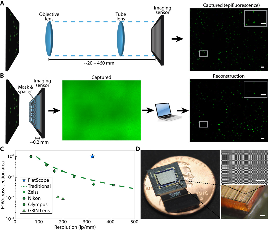
Fig. 1 Traditional microscope versus FlatScope. (A) Traditional microscopes capture the scene through an objective and tube lens (~20 to 460 mm), resulting in a quality image directly on the imaging sensor. (B) FlatScope captures the scene through an amplitude mask and spacer (~0.2 mm) and computationally reconstructs the image. Scale bars, 100 μm (inset, 50 μm). (C) Comparison of form factor and resolution for traditional lensed research microscopes, GRIN lens microscope, and FlatScope. FlatScope achieves high-resolution imaging while maintaining a large ratio of FOV relative to the cross-sectional area of the device (see Materials and Methods for elaboration). Microscope objectives are Olympus MPlanFL N (1.25×/2.5×/5×, NA = 0.04/0.08/0.15), Nikon Apochromat (1×/2×/4×, NA = 0.04/0.1/0.2), and Zeiss Fluar (2.5×/5×, NA = 0.12/0.25). (D) FlatScope prototype (shown without absorptive filter). Scale bars, 100 μm.

Science Advances (2017)

Single-frame 3D fluorescence microscopy with ultraminiature lensless FlatScope

Jesse K Adams, Vivek Boominathan, Benjamin W Avants, Daniel G Vercosa, Fan Ye, Richard G Baraniuk, Jacob T Robinson, Ashok Veeraraghavan 

Thrust 3 Publications

PATHS-UP Members

Texas A&M University
UCLA
Rice University
Florida International University

Evaluation Partner

Arizona State University

Funded By

Copyright © 2025 · Texas A&M Engineering Experiment Station · All Rights Reserved

Accessibility • Site Links & Policies • Privacy Policy • Website Feedback • Texas A&M University